Thursday, February 2, 2012
Virtual Leech Lab!~ All about Neurophysiology
So in Science(or maybe only in science with Mr. Ludwig) you don't always do conduct a lab in person. To learn about Neurophysiology our class did an online virtual lab that involved leeches.(I'm actually kind of glad it was an online lab!)
Objective:
In this lab we wanted to record electrical activities of individual neurons while delivering mechanical stimulus to the attached skin and inject fluorescent dyes into the neurons to visualize their morphology. We also wanted to identify the neurons based on the morphology and the response to stimuli, comparing them to previously published results.
(Some quick definitions to help you understand this experiment!
-Neurophysiology: A study of physical and chemical processes of neurons
-Neuron: A nerve cell
-Morphology: The form and structure of any organism or any of it's parts. ~In this experiment it is the neuron.
Materials:-Feather: This is used to give the leech skin a very gentle touch stimulation
-Probe: This is a blunt metal rod attached to a wooden handle and in this experiment ou use it to lift tissue, and to push the skin as a stimulus.
-Forceps: These are fine forceps for very fine manipulations.(also known as Dumont #5)
-Scissors: These need to be good dissecting angled scissors. For this experiment they cut open the body wall.
-Pins: These are stainless steel dissecting pins for pinning tissue to a dissection dish.
-Scalpel: For this experiment you need disposable scalpel blades because they are better for microsurgery.
-Dissection Tray: This is a tray that is half-filled with hard wax so that you can stick pins into it to secure the leech for dissection.
-Leech Tank: For this experiment it is best to keep leeches in pond water.
-20% Ethanol: This is used to anesthetize the leech. (Time for the first FUN FACT of the semester!: 20% ethanol is humane and stops the leech from moving, which makes it easier to pin it down.
-Leech Tongs: These are basically gross anatomy forceps and are used to pick up the leech.
-Dissection Microscope: This is a special microscope designed for dissections. It is basically a high quality high power magnifying glass.
-Micromanipulator: This is a device that is used to position items with sub- micrometer precision in three dimensions. In this experiment we mount our electrode on it to guide it accurately to a neuron.
-Oscilloscope: This is basically a sophisticated voltmeter. [On the screen you see a real time display of voltage(vertical) plotted against time(horizontal)]
-Leech: The thing we are dissecting!
Procedure:
1. First we caught the leech and anesthetized it in 20% ethanol solution.
2. We pinned the leech to the dissection dorsal(the top) side up. We stretched the animal out in the process.
3. Using the scissors we cut the skin along the mid-line on the dorsal side. We were very careful not to damage the deep structures. Then we used the forceps to carefully peel apart the skin and pin down each side of skin. (FUN FACT: DOing this exposes the innards of the leech, including the digestive, excretory, and reproductive organs!)
4. Next we carefully removed the gut and the other internal structures so we could see the ventral nerve cord.
5. We moved the leech under the Dissection microscope so we could look at the ganglia of the nervous system. (a collection of neuronal cell bodies. Using the scalpel we cut a window along the sinus(circulation system of a leech) under a ganglion. We made sure not to do any damage to the nerves.
6. Using the scalpel we cut a section of ganglion out of the leech making two parallel cuts that were perpendicular to the anterior-posterior axis. Then using forceps we flipped the piece of skin so that the outer skin was facing up and pinned the skin down.
7. Under the dissection microscope we cut the sinus with an ultra fine scalpel and using fine forceps we carefully peeled apart the sinus to expose the ganglion. We could then see individual cells under the microscope.
8. We moved the skin under the Micromanipulator to find the cells. We used the electrode to gain control of it and moved the electrode any place over the ganglion to stimulate penetrating the cell. (this is actually much harder in real life) We watched the oscilloscope so we could see when we had found a cell. The oscilloscope turned the signals from the cell into a sound wave display. Then we used a feather, probe, and forceps to see if the cell would respond to see any stimulus. When the cell showed a spike it responded in firing an action potential. Then we compared our data to the data in the atlas. Then we dyed the cells with the fluorescent dye to see where the different cells were located.
9. Next we used the UV Switch to see the dye. This let us see the structure of the cells.
10. At the end we used the electrophysiological and neuronanatomical data to identify each cell. After we identified one cell we went back and identified the other 4 cells using the same process.
Analyzing Data:
All together I identified 5 cells. I could tell what was each cell by how it responded to the different stimulus.
The atlas above is what I used to compare my results to. In this experiment I learned how the different cell types react to the different stimulus. For example cell type N only reacted to the forceps while cell type P only reacted to the probe. Some cells, like cell type R, fire action potentials without any stimulus affecting them and some cells, like cell type X, don't react to any stimulus at all. Cell type T responded differently to each of the different stimulus.
Conclusion:
This experiment taught me a lot about the body. There are so many different types of cells in one small ganglion. (a collection of neuronal cell bodies). All the cells are different. They each respond differently to the different stimuli. This virtual lab helped me understand the different types of cells and how they are different.
If you would like to try this online virtual lab follow this link! (I suggest it!)
http://www.hhmi.org/biointeractive/vlabs/neurophysiology/index2startlab.html
Concussions and the Brain
This week I got together with Madison and Ali. We made a prezi about Concussions and how they affect the Nervous System. Then we had to give a presentation in front of the class and make quiz questions to put on a quiz! We'll here is our prezi!
See if you can answer the quiz questions!
-Name two common symptoms of a concussion? headache, amnesia, confusion
-How long can symptoms of a concussion last? can be as long as weeks or months depending on the severity
-How do concussions happen: the brain knocks around the skull and it bruises breaking blood vessels and affecting nervous system
-True or False? Depending on the severity of the concussion a concussion can actually change the way the brain works......TRUE
-True or False? A person can obtain a concussion only when they hit their head.....FALSE~anything hit that can make the brain move
See if you can answer the quiz questions!
-Name two common symptoms of a concussion? headache, amnesia, confusion
-How long can symptoms of a concussion last? can be as long as weeks or months depending on the severity
-How do concussions happen: the brain knocks around the skull and it bruises breaking blood vessels and affecting nervous system
-True or False? Depending on the severity of the concussion a concussion can actually change the way the brain works......TRUE
-True or False? A person can obtain a concussion only when they hit their head.....FALSE~anything hit that can make the brain move
Concussions on Prezi
Tuesday, December 20, 2011
Pituitary Gigantism and Acromegaly~Research
Recently I watched a video on Edmodo about Pituitary Gigantism and Acromegaly. Pituitary Gigantism is caused when there is a tumor on your pituitary gland that triggers a surge that causes excess growth hormones. This is what leads to the huge size of Pituitary Giants.
The excess growth hormones produce large internal organs. Pituitary giants have organs that are two times the normal volume of regular organs. Their organs are the right size for their bodies though becuase their bodies are two times the normal volume also.
The only organ that is the same size as the average human is the brain. This is because the excess growth hormaone makes the walls of the skull grow thicker. This gives the brain extra protection but it makes the cavity stay the same size. This prevents the brain from growing any bigger then the size of an average human.
Usually when you see a Pituitary Giant you associate their size with a lot of strenth. They do have a lot of muscle mass and have big muscles but giants are not as strong as they look. The excess growth hormones also cause an excess of salt in the muscle tissue. The salt makes the muscles swell up with water making the muscles bigger but not stronger. Their muscle development is also affected by the way their bones grew. In their extreme growth sometimes the nerves on one side of a muscle grow faster then the other side. This cause the bone to curve and it causes the muscles to be like rubber bands that are to long and to slack. In the end the muscles are weaker. Pituitary giants have bodies that are "wired" differently then the average person and this can also affect their muscle power. The excess of growth hormone causes problems with the peripheral nervous system. This makes a change on the way the skeletal muscles move. Their peripheral nerves span out from the centeral nerves of the brain and the spinal column. These nerves cause electrical impulses at very very high speeds(more than 200 miles per hour!) that cause the muscles to contract. These peripheral nerves are made of long cells that are surrounded by a protective insulation, think of electrical wires. In pituitary gigantism the excess growth hormone causes the inflimation to swell up. This restricts the movement of the nerve impulses and restricts the movement of the skeletal muscles. Their muscles lose strength and don't work like they usually would. Regular activities, like walking, can be tiring because of the excess growth hormone that gives them their size also makes them more tired.
Sometimes these hormones never stop. Sometimes surgery, radiation, therapy,and drugs can be used to get the excess growth hormone under control and stop the growing in pituitary giants. If the growth hormone doesn't get under control their bones still grow. The bones don't grow taller though. The bones grow thicker. This is called Acromegalic Gigantism. Once the body can't get any taller the bones start growing larger and becomemore prominent. The obvious signs of this are usually in the skull. Acromegaly causes a more prominent forehead, makes the eyebrow bones bulge, and causes more prominent cheekbones. It also causes the lower jaw to grow and push the bottom teeth out past the upper teeth. It can also cause a broader nose, thicker lips, and a larger tounge with a lower voice. Many giants have a male sounding voice. They can become winded very easily.
After doing further research I learned that most cases of Pituitary Gigantism can be helped with pituitary surgery. This surgery is known to usually cure most cases. The next choice of treatment for most people is medication. The most effective medications known are somatostatin analogs. These reduce the release of growth hormone. There is also a medication known as Dopamine agonists. These have not been proven as effective the other though. Sometimes radiation has been used as cures.
To see the video that taught me A LOT follow this link:
http://www.youtube.com/watch?v=Ebhf1qKVA9A
To read the article I read follow this link:
http://health.nytimes.com/health/guides/disease/gigantism/overview.html
The excess growth hormones produce large internal organs. Pituitary giants have organs that are two times the normal volume of regular organs. Their organs are the right size for their bodies though becuase their bodies are two times the normal volume also.
The only organ that is the same size as the average human is the brain. This is because the excess growth hormaone makes the walls of the skull grow thicker. This gives the brain extra protection but it makes the cavity stay the same size. This prevents the brain from growing any bigger then the size of an average human.
Usually when you see a Pituitary Giant you associate their size with a lot of strenth. They do have a lot of muscle mass and have big muscles but giants are not as strong as they look. The excess growth hormones also cause an excess of salt in the muscle tissue. The salt makes the muscles swell up with water making the muscles bigger but not stronger. Their muscle development is also affected by the way their bones grew. In their extreme growth sometimes the nerves on one side of a muscle grow faster then the other side. This cause the bone to curve and it causes the muscles to be like rubber bands that are to long and to slack. In the end the muscles are weaker. Pituitary giants have bodies that are "wired" differently then the average person and this can also affect their muscle power. The excess of growth hormone causes problems with the peripheral nervous system. This makes a change on the way the skeletal muscles move. Their peripheral nerves span out from the centeral nerves of the brain and the spinal column. These nerves cause electrical impulses at very very high speeds(more than 200 miles per hour!) that cause the muscles to contract. These peripheral nerves are made of long cells that are surrounded by a protective insulation, think of electrical wires. In pituitary gigantism the excess growth hormone causes the inflimation to swell up. This restricts the movement of the nerve impulses and restricts the movement of the skeletal muscles. Their muscles lose strength and don't work like they usually would. Regular activities, like walking, can be tiring because of the excess growth hormone that gives them their size also makes them more tired.
Sometimes these hormones never stop. Sometimes surgery, radiation, therapy,and drugs can be used to get the excess growth hormone under control and stop the growing in pituitary giants. If the growth hormone doesn't get under control their bones still grow. The bones don't grow taller though. The bones grow thicker. This is called Acromegalic Gigantism. Once the body can't get any taller the bones start growing larger and becomemore prominent. The obvious signs of this are usually in the skull. Acromegaly causes a more prominent forehead, makes the eyebrow bones bulge, and causes more prominent cheekbones. It also causes the lower jaw to grow and push the bottom teeth out past the upper teeth. It can also cause a broader nose, thicker lips, and a larger tounge with a lower voice. Many giants have a male sounding voice. They can become winded very easily.
After doing further research I learned that most cases of Pituitary Gigantism can be helped with pituitary surgery. This surgery is known to usually cure most cases. The next choice of treatment for most people is medication. The most effective medications known are somatostatin analogs. These reduce the release of growth hormone. There is also a medication known as Dopamine agonists. These have not been proven as effective the other though. Sometimes radiation has been used as cures.
To see the video that taught me A LOT follow this link:
http://www.youtube.com/watch?v=Ebhf1qKVA9A
To read the article I read follow this link:
http://health.nytimes.com/health/guides/disease/gigantism/overview.html
Monday, December 19, 2011
EMG Lab
To start off our unit on the Muscular System we had an EMG lab. EMG is a graphical recording of electrical activity within the muscles. When the muscles are activated by nerves it results in changes in ion flow across cell membranes. This generates electrical activity. In this lab we were trying to create a graph that represented the electrical activity of a muscle and how much energy was used when a muscle would contract. So for our experiment we did a chewing activity to evaluate the muscles in the jaw when chewing different types of food.
Hypothesis:
If our group tests the muscles in the jaw while someone chews different foods then the harder foods will make the muscles work more and will generate more electrical activity on the graph because your muscles in the jaw work harder to chew harder foods then they do to chew softer foods.
Materials:
Eater (to eat the different foods)
Different types of food
EKG probe and electrode tabs
Experiment:
After setting up the computer we hooked our "Eater" Sierra up to the probes. We put a probe on her upper jaw and lower jaw. First we set up a baseline by having Sierra clench her jaw 30 seconds. Then we gave Sierra different foods that were varying hardness to chew slowly. Sierra would rest her jaw for 5 seconds then start chewing the food until swallowing. The clenching data was used to compare to the other foods that were chewed. The foods Sierra tried were barbeque chips, a carrot, chocolate chip cookies, a banana, celery, beef jerky, and pudding. We also had her drink Dr. Pepper.
DATA:
Data Analysis:
From the data we collected you can see that the different foods caused higher and lower electrical activity. The clenching of the jaw we used as average jaw activity at 0.5 mV. The food that caused the highest amount of electrical activity was the pudding at 2.2 mV. We think this is because when people chew they normally don't open their mouth very much but when people eat pudding they tend to open their mouth wider. The food that caused the lowest amount of electrical activity was the chocolate chip cookie at 0.3 mV. Along with the cookie the Dr. Pepper also created electrical activity at 0.3 mV. The other foods rated everywhere between these two. The celery came the closest to the baseline at 0.6 mV.
Conclusion:
After our experiment we concluded that our hypothesis was not always true. Foods that are harder tended to make our jaw work harder but it is not always the case. Sometimes the way your mouth chews depends on the texture of the food. Something sticky and creamy like pudding can make your mouth open wider which causes more electrical activity. Our graph and analysis prove this.
Reflection:
I really enjoyed this lab. It was a really good way to start off the muscular system! I liked this lab because it made me think about muscles that I never really think about. I never thought about the jaw working in that way. Our group worked very well together to finish our project. I really enjoyed this lab because of how much fun we had doing it. It was also a good idea to call the lab a "Potluck!" I think our experiment was a great success and a lot of fun!
Thursday, December 15, 2011
Muscle Anatomy
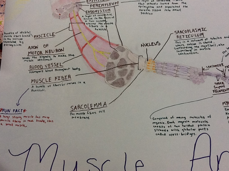
To learn about the Musclular System our class split into different groups and picked one of three things to research: Muscle Anatomy, Sliding Filament Theory(FUN FACT: This is how we think muscles contract!), and Neuromuscular Junction.
Well Ali, Madison, and I all got together to create a poster about the structure of a muscle (muscle anatomy). We all worked together to research the muse anatomy(on our IPads by the way!) Something I learned while making this poster....I have a little talent in drawing! Well I hope you enjoy our poster!
Monday, December 5, 2011
Skeletal System
So I attempted another prezi! I tried to make this one a little more interesting and use different things like colors and turning my words. Hope you enjoy!
Here's the link to my prezi:
I wanted to take this time to do a little self anaylsis. I have been really proud of myself this quarter. For the most part I have stayed caught up with my blogs. I didn't let logs pile up and have to rush getting them done at the end of the semester. The most I got behind was getting this blog done. I have also been studying for my quizzes this year, unlike last year in Biology. I have not gotten under a 95% on any of my quizzes this year. I am getting better at using Anatomy class time for Anatomy work. I can still be better at using my time better but I am doing better then I was last semester.( this is a tongue twister I know!) My goal is to get an A for this semester. I think this is possible because I am understanding things better. The notes on the Ipad have really helped me! I feel like it gives me a direction on what information I need to provide. Last year I was always nervous that I wasn't writing the right information that Mr. Ludwig wanted. LJHS something to think about, I think you should supply Ipad's for all of Mr. Ludwig's classes. Just something for our school to think about....
Here's the link to my prezi:
I wanted to take this time to do a little self anaylsis. I have been really proud of myself this quarter. For the most part I have stayed caught up with my blogs. I didn't let logs pile up and have to rush getting them done at the end of the semester. The most I got behind was getting this blog done. I have also been studying for my quizzes this year, unlike last year in Biology. I have not gotten under a 95% on any of my quizzes this year. I am getting better at using Anatomy class time for Anatomy work. I can still be better at using my time better but I am doing better then I was last semester.( this is a tongue twister I know!) My goal is to get an A for this semester. I think this is possible because I am understanding things better. The notes on the Ipad have really helped me! I feel like it gives me a direction on what information I need to provide. Last year I was always nervous that I wasn't writing the right information that Mr. Ludwig wanted. LJHS something to think about, I think you should supply Ipad's for all of Mr. Ludwig's classes. Just something for our school to think about....
Friday, November 4, 2011
Integumentary System
So we continue or way through the study of our body. Next we come to the Integumentary system!
So when you hear someone talking about the integumentary system they are talking about your skin. Our skin is made up of three major regions:
-Epidermis: This is the outermost superficial region.
-Dermis: This is the middle region.
-Hypodermis: This is the deepest region. It is also referred to as the superficial fascia.
The Epidermis is made of keratinized stratified squamos epithelium. It consists of four distinct cell types and around four or five layers.
The cell types in the Epidermis include:
-Keratinocytes: These produce the fibrous protein keratin
-Melanocytes: These produce brown piment melanin
-Langerhans' cells: These are epidermal macrophages that help activate the immune system.
-Merkel Cells: These function as tough receptors in association with sensory nerve endings.
The outer protion of the skin is exposed to the external environment. It functions in protection.
The epidermis is split into different layers!
The first layer is the Stratum Basale or the Basal Layer. This is the deepest layer and is firmly attached to the dermis. The basal layer consists of a single row of the youngest keratinocytes. These cells undergo rapid division. (FUN FACT: It's alternate name is Stratum Germinativum!)
The next layer of the Epidermis is the Stratum Spinosum. It is also called the Prickly layer. The cells contain a weblike system of intermediate filaments attached to demosomes. Melanin granules and Langerhans' cells are very abundant in this layer.
Next comes the Stratum Granulosum, also known as the granular layer. This layer has around three to five cell layers, where drastic changes in keratinocyte appearance occurs. It is a thin layer where keratohyaline and lamellated granules accumulate.
The next layer is know as the Stratum Lucidum, or also called the clear layer. This layer is a thin, transparent band superficial to the stratum granulosum. It has a few rows of flat, dead keratinocytes. (FUN FACT: This layer is only present in thick skin!)
The top, or outermost layer of keratinized cells, layer of the epidermis is the Stratum Corneum, or the Horny layer. This layer accounts for three quarters of the epidermal thickness. This layer has three different functions; Waterproofing, protection from abrasion and penetration, and rendering the body relatively insensitive to biological, chemical, and physical assaults.
The next layer of skin is the Dermis.
This is the second major skin region. it contains strong, flexible connective tissue. The cell types in the dermis can include fibroblasts, macrophages, and occasionally mast cells and white blood cells.
The Dermis is split into two layers!
The Papillary layer is areolar connective tissue with collagen and elastic fibers. This layer's superior surface has peglike projections that are called dermal papillae. The dermal papillae are made of capillary loops, Meissner's corpuscles, and free nerve endings.
The next layer in the dermis is the Reticular layer. This layer makes up around 80% of the thickness of the skin. The collagen fibers found in this layer add strength and resiliency to the skin, The elastin fibers stretch-recoil properties.
The next major layer of the skin is the Hypodermis. This is the subcutaneous layer deep to the skin. It is made of adipose and areolar connective tissue.
Everyone has different skin colors. We have three pigments that contribute to what color skin we have.
-Melanin: This is a yellow to reddish-brown to black pigment. It is responsible for dark skin colors. Freckles and pigmented moles result from local accumulations of melanin.
-Carotene: This is a yellow to orange pigment. It is most obvious in the palms and soles of the feet.
-Hemoglobin: This is a reddish pigment responsible for the pinkish hue in the skin.
Now everybody sweats but did you know we have different types of sweat glands that prevent the body from overheating.
-Eccrine Sweat Glands are found in the palms, soles of the feet, and the forehead.
-Apocrine Sweat Glands are found in axillary and anogenital areas.
-Ceruminous Glands are modified apocrine glands in the external ear canal that secrete cerumen.
-Mammary Glands are specialized sweat glands that secrete milk.
Our body also has another kind of gland: Sebaceous Glands. These are simple alveolar glands that are found all over the body. They secrete an oily secretion called sebum. They also soften the skin when its stimulated by hormones. (FUN FACT: You can blame these little guys for acne!)
FUN FACT: Have you ever wondered about your nails!? Well nails are a scalelike modification of the epidermis on the distal, dorsal surface of the fingers and toes!
Most girls love to have great hair! Some guys too! But do you really know everything there is to know about hair?!
Your hair is filamentous strands of dead keratinized cells that are produced by hair follicles. It has hard keratin! (FUN FACT: Hard keratin is tougher and more durable than soft keratin of the skin!) Your hair is made up of the shaft projecting from the skin and the root embedded in the skin. It also has a core called the medulla, a cortex, and an outermost cuticle. Your hair is pigmented by melanocytes at the base of the hair. (in other words that how you get your hair color!) You hair also has different functions!
-It helps to maintain warmth
-It alerts your body when insects are on your skin
-It guards the scalp against physical trauma, heat loss, and sunlight.
You can find hair anywhere on your body except for palms, soles, lips, nipples, and portions of the external genitalia.
Your hair grows from hair follicles on your scalp. A hair follicle is a root sheath extending from the epidermal surface into the dermis. The deep end is expanded forming a hair bulb. Wrapped around each hair bulb is a knot of sensory nerve endings, also known as a root hair plexus. If you bend a hair it can stimulate these endings. Our hairs act as sensitive touch receptors.
Our Integumentary System has different functions as a whole. Our skin is there for protection. It is a chemical, physical, and mechanical barrier. It also regulates our body temperatures. It can do this by Dilation, cooling, and constriction, warming, of the dermal vessels. Our exoreceptors can also sense touch and pain, which is known as the cutaneous sensation. Our skin also has metabolic functions and synthesis of vitamin D in dermal blood vessels. Our skin blood vessels store up to 5% of the body's blood volume. Our skin also goes through excretion where limited amount of nitrogenous wastes are eliminated from the body through sweating.
Our skin can get three major types of skin cancer; Basal cell carcinoma, squamos cell carcinoma, and melanoma. Melanoma, or the cancer of melanocytes is the most dangerous type of skin cancer. This is because Melanoma is highly metastic and is resistant to chemotherapy. You can tell if someone has melanoma by using the ABCD rule.
-A: Asymmetry; The two sides of the pigmented area do not match
-B: Border; It is irregular and exhibits indentations.
-C: Color; The pigmented area is black, brown, tan, and can sometimes be even red or blue.
-D: Diameter; The diameter is larger than 6mm, or the size of a pencil eraser.
This cancer is treated by wide surgical excision accompanied by immunotherapy. The chance of surviving is very poor if the lesion is over 4 mm thick.
Now its time to talk about burns.
In first-degree burns only the epidermis is damaged. The symptoms include localized redness, swelling, and pain.
In second-degree burns the epidermis and upper regions the dermis are damaged. The symptoms mimic first degree burns but blisters also appear.
In third-degree burns the entire thickness of the skin is damaged. The burned are appears gray-white, cherry red, or black. Your nerve endings are destroyed so there is no initial edema or pain.
The Rule of nines estimates the severity of burns. Your burns are considered critical if:
-Over 25% of the body has second-degree burns
-Over 10% of the body has third-degree burns.
-There are third-degree burns on face, hands, or feet.
Now I know what you're thinking...OH MY GOSH THAT WAS LONG! I bet you never thought there was that much involved in your skin. Well trust me there's even more information to know that I don't even know! Well I hope you now know more about our skin then you did before.
Be looking for my next very very very long post about our Skeletal System!!
So when you hear someone talking about the integumentary system they are talking about your skin. Our skin is made up of three major regions:
-Epidermis: This is the outermost superficial region.
-Dermis: This is the middle region.
-Hypodermis: This is the deepest region. It is also referred to as the superficial fascia.
The Epidermis is made of keratinized stratified squamos epithelium. It consists of four distinct cell types and around four or five layers.
The cell types in the Epidermis include:
-Keratinocytes: These produce the fibrous protein keratin
-Melanocytes: These produce brown piment melanin
-Langerhans' cells: These are epidermal macrophages that help activate the immune system.
-Merkel Cells: These function as tough receptors in association with sensory nerve endings.
The outer protion of the skin is exposed to the external environment. It functions in protection.
The epidermis is split into different layers!
The first layer is the Stratum Basale or the Basal Layer. This is the deepest layer and is firmly attached to the dermis. The basal layer consists of a single row of the youngest keratinocytes. These cells undergo rapid division. (FUN FACT: It's alternate name is Stratum Germinativum!)
The next layer of the Epidermis is the Stratum Spinosum. It is also called the Prickly layer. The cells contain a weblike system of intermediate filaments attached to demosomes. Melanin granules and Langerhans' cells are very abundant in this layer.
Next comes the Stratum Granulosum, also known as the granular layer. This layer has around three to five cell layers, where drastic changes in keratinocyte appearance occurs. It is a thin layer where keratohyaline and lamellated granules accumulate.
The next layer is know as the Stratum Lucidum, or also called the clear layer. This layer is a thin, transparent band superficial to the stratum granulosum. It has a few rows of flat, dead keratinocytes. (FUN FACT: This layer is only present in thick skin!)
The top, or outermost layer of keratinized cells, layer of the epidermis is the Stratum Corneum, or the Horny layer. This layer accounts for three quarters of the epidermal thickness. This layer has three different functions; Waterproofing, protection from abrasion and penetration, and rendering the body relatively insensitive to biological, chemical, and physical assaults.
The next layer of skin is the Dermis.
This is the second major skin region. it contains strong, flexible connective tissue. The cell types in the dermis can include fibroblasts, macrophages, and occasionally mast cells and white blood cells.
The Dermis is split into two layers!
The Papillary layer is areolar connective tissue with collagen and elastic fibers. This layer's superior surface has peglike projections that are called dermal papillae. The dermal papillae are made of capillary loops, Meissner's corpuscles, and free nerve endings.
The next layer in the dermis is the Reticular layer. This layer makes up around 80% of the thickness of the skin. The collagen fibers found in this layer add strength and resiliency to the skin, The elastin fibers stretch-recoil properties.
The next major layer of the skin is the Hypodermis. This is the subcutaneous layer deep to the skin. It is made of adipose and areolar connective tissue.
Everyone has different skin colors. We have three pigments that contribute to what color skin we have.
-Melanin: This is a yellow to reddish-brown to black pigment. It is responsible for dark skin colors. Freckles and pigmented moles result from local accumulations of melanin.
-Carotene: This is a yellow to orange pigment. It is most obvious in the palms and soles of the feet.
-Hemoglobin: This is a reddish pigment responsible for the pinkish hue in the skin.
Now everybody sweats but did you know we have different types of sweat glands that prevent the body from overheating.
-Eccrine Sweat Glands are found in the palms, soles of the feet, and the forehead.
-Apocrine Sweat Glands are found in axillary and anogenital areas.
-Ceruminous Glands are modified apocrine glands in the external ear canal that secrete cerumen.
-Mammary Glands are specialized sweat glands that secrete milk.
Our body also has another kind of gland: Sebaceous Glands. These are simple alveolar glands that are found all over the body. They secrete an oily secretion called sebum. They also soften the skin when its stimulated by hormones. (FUN FACT: You can blame these little guys for acne!)
FUN FACT: Have you ever wondered about your nails!? Well nails are a scalelike modification of the epidermis on the distal, dorsal surface of the fingers and toes!
Most girls love to have great hair! Some guys too! But do you really know everything there is to know about hair?!
Your hair is filamentous strands of dead keratinized cells that are produced by hair follicles. It has hard keratin! (FUN FACT: Hard keratin is tougher and more durable than soft keratin of the skin!) Your hair is made up of the shaft projecting from the skin and the root embedded in the skin. It also has a core called the medulla, a cortex, and an outermost cuticle. Your hair is pigmented by melanocytes at the base of the hair. (in other words that how you get your hair color!) You hair also has different functions!
-It helps to maintain warmth
-It alerts your body when insects are on your skin
-It guards the scalp against physical trauma, heat loss, and sunlight.
You can find hair anywhere on your body except for palms, soles, lips, nipples, and portions of the external genitalia.
Your hair grows from hair follicles on your scalp. A hair follicle is a root sheath extending from the epidermal surface into the dermis. The deep end is expanded forming a hair bulb. Wrapped around each hair bulb is a knot of sensory nerve endings, also known as a root hair plexus. If you bend a hair it can stimulate these endings. Our hairs act as sensitive touch receptors.
Our Integumentary System has different functions as a whole. Our skin is there for protection. It is a chemical, physical, and mechanical barrier. It also regulates our body temperatures. It can do this by Dilation, cooling, and constriction, warming, of the dermal vessels. Our exoreceptors can also sense touch and pain, which is known as the cutaneous sensation. Our skin also has metabolic functions and synthesis of vitamin D in dermal blood vessels. Our skin blood vessels store up to 5% of the body's blood volume. Our skin also goes through excretion where limited amount of nitrogenous wastes are eliminated from the body through sweating.
Our skin can get three major types of skin cancer; Basal cell carcinoma, squamos cell carcinoma, and melanoma. Melanoma, or the cancer of melanocytes is the most dangerous type of skin cancer. This is because Melanoma is highly metastic and is resistant to chemotherapy. You can tell if someone has melanoma by using the ABCD rule.
-A: Asymmetry; The two sides of the pigmented area do not match
-B: Border; It is irregular and exhibits indentations.
-C: Color; The pigmented area is black, brown, tan, and can sometimes be even red or blue.
-D: Diameter; The diameter is larger than 6mm, or the size of a pencil eraser.
This cancer is treated by wide surgical excision accompanied by immunotherapy. The chance of surviving is very poor if the lesion is over 4 mm thick.
Now its time to talk about burns.
In first-degree burns only the epidermis is damaged. The symptoms include localized redness, swelling, and pain.
In second-degree burns the epidermis and upper regions the dermis are damaged. The symptoms mimic first degree burns but blisters also appear.
In third-degree burns the entire thickness of the skin is damaged. The burned are appears gray-white, cherry red, or black. Your nerve endings are destroyed so there is no initial edema or pain.
The Rule of nines estimates the severity of burns. Your burns are considered critical if:
-Over 25% of the body has second-degree burns
-Over 10% of the body has third-degree burns.
-There are third-degree burns on face, hands, or feet.
Now I know what you're thinking...OH MY GOSH THAT WAS LONG! I bet you never thought there was that much involved in your skin. Well trust me there's even more information to know that I don't even know! Well I hope you now know more about our skin then you did before.
Be looking for my next very very very long post about our Skeletal System!!
Subscribe to:
Posts (Atom)